Small angle X-ray scattering (SAXS) and small angle neutron scattering (SANS) of viral genomes
Despite the structure of packaged viral DNA being central for ejection and consequentially infectivity of the virus, the detailed understanding of the packing structure of DNA inside the viral capsid. SAXS and SANS allow investigation of the structure of DNA filled capsids in solution in a wide range of environmental conditions. SAXS and SANS are powerful techniques that provides detailed structural information on the virally encapsidated genome. Since capsid protein structure and DNA have different scattering profiles, with DNA having a short-range hexagonally ordered structure, we can observe the scattering peak for the packaged DNA in viral capsid. Our preliminary SAXS data from both in house bench type SAXS and synchrotron SAXS (at Argonne National Lab) clearly demonstrate the presence of such a well-resolved DNA-DNA diffraction peak. The position of the DNA diffraction peak corresponds to the DNA inter-axial spacing, which describes the short-range genome ordering. At the same time, the width and the integrated area of the DNA diffraction peak, describes the long-range genome ordering. If the DNA inside the capsid becomes less long-range ordered, the peak width is increasing, while its area is decreasing. If the genome is completely disordered, the DNA diffraction peak disappears. We use solution SAXS to investigate the structure and long-range structural phase-transition of the encapsidated genomes under variable environmental parameters.
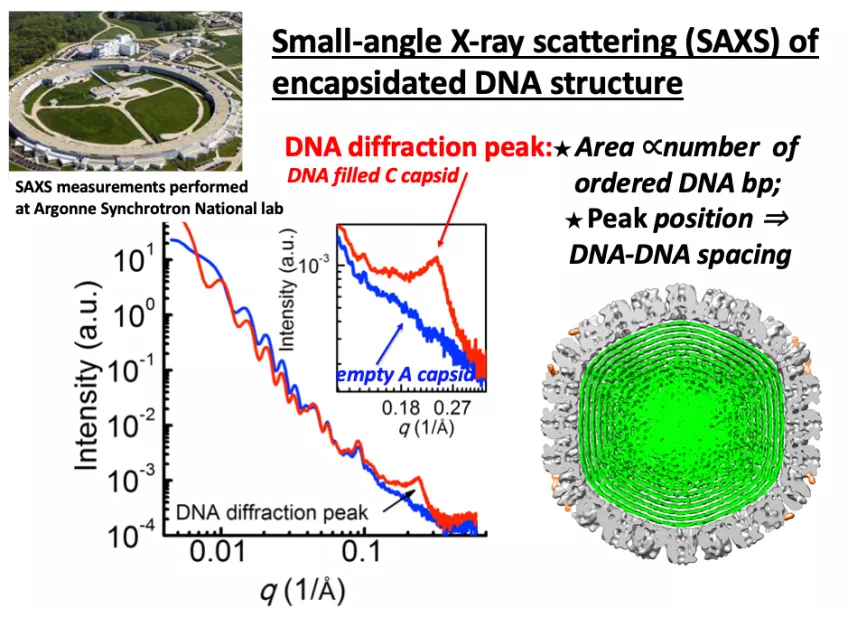
Figure: Solution SAXS analysis of intracapsid DNA structure for HSV-1. (a) Background subtracted SAXS data, I(q) versus q, for empty HSV-1 A-capsid (blue curve) and DNA-filled C-capsid (red curve). The inset figure shows a zoomed-in region of the DNA diffraction peak from C-capsid. AU, arbitrary units.
MAX IV Laboratory
European Spallation Source