Cryo-Electron Microscopy
We use cryo electron microscopy (cryo-EM) and image reconstruction to examine the virion structure at subnanometer resolution. We also analyze structure and density of viral DNA packaged in a capsid.
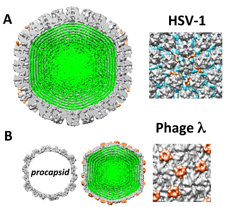
Figure: Cutaway views from cryo-EM reconstructions of (A) HSV-1 C-capsid and (B) phage lambda procapsid and mature DNA-filled lambda capsid. HSV-1 and lambda phage capsids are shown to scale. Owing to the icosahedral symmetry imposed during the reconstruction, concentrically packed DNA within the capsid becomes shells of density (green).